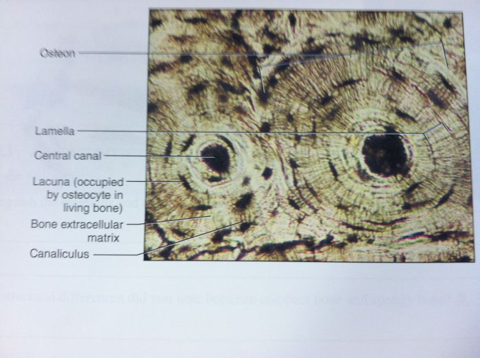
On the photomicrograph of bone below, we delve into the intricate histological structure of bone tissue, exploring its cellular components, organization, and clinical significance. This microscopic examination unveils the secrets of bone’s remarkable strength, adaptability, and regenerative capabilities.
The photomicrograph captures the essence of bone’s architecture, showcasing osteocytes nestled within lacunae, interconnected by a network of canaliculi. The organized arrangement of lamellae and Haversian systems reveals the meticulous design that underlies bone’s mechanical properties.
Histology of Bone: On The Photomicrograph Of Bone Below
Bone is a mineralized connective tissue that forms the skeletal system. It provides support, protection, and movement for the body. Bone is composed of cells, extracellular matrix, and mineral crystals.
The histological structure of bone is complex and highly organized. It consists of several different cell types, including osteoblasts, osteocytes, and osteoclasts. These cells are responsible for the formation, maintenance, and resorption of bone tissue.
Cells of Bone
- Osteoblastsare bone-forming cells that secrete the organic matrix of bone, which is composed of collagen and other proteins. Osteoblasts are located on the surface of bone and are responsible for the growth and repair of bone tissue.
- Osteocytesare mature bone cells that are embedded within the mineralized matrix of bone. Osteocytes are responsible for maintaining the bone matrix and regulating the exchange of nutrients and waste products between the blood and the bone.
- Osteoclastsare bone-resorbing cells that are responsible for breaking down and removing bone tissue. Osteoclasts are located on the surface of bone and secrete acids and enzymes that dissolve the mineralized matrix of bone.
Organization of Bone Tissue
Bone tissue is organized into lamellae, which are thin layers of mineralized matrix. Lamellae are arranged in concentric circles around a central canal, which contains blood vessels and nerves. The central canal is surrounded by osteocytes, which are located within lacunae, or small cavities, in the mineralized matrix.
The lamellae are further organized into Haversian systems, which are cylindrical units of bone tissue. Haversian systems are composed of several lamellae, which are arranged around a central canal. The central canal contains blood vessels and nerves, which supply the osteocytes with nutrients and oxygen.
Photomicrograph of Bone

A photomicrograph of bone tissue provides a detailed view of the microscopic structure of bone. It can reveal the different types of cells, the arrangement of the extracellular matrix, and the organization of the tissue into functional units.
Bone Structure
The photomicrograph shows the following structures:
- Osteocytes:These are mature bone cells that are embedded within the mineralized matrix of the bone. They are located in small cavities called lacunae.
- Lacunae:These are small cavities that house the osteocytes. They are connected to each other by a network of small channels called canaliculi.
- Canaliculi:These are small channels that connect the lacunae to each other and to the central canal of the Haversian canal. They allow for the exchange of nutrients and waste products between the osteocytes and the blood vessels in the Haversian canal.
- Haversian canals:These are larger channels that run through the bone tissue. They contain blood vessels and nerves that supply the osteocytes with nutrients and oxygen.
The photomicrograph can be used to study the histology of bone, which is the study of the microscopic structure of bone tissue. It can provide information about the health and condition of the bone, as well as the effects of various factors, such as age, disease, and treatment.
Analysis of Bone Photomicrograph

The photomicrograph of bone reveals a complex and highly organized tissue. The cells, structures, and matrix of bone work together to provide strength, support, and protection for the body.
Types of Cells in Bone, On the photomicrograph of bone below
- Osteoblasts: These cells are responsible for the formation of new bone tissue. They secrete a matrix of collagen and other proteins, which mineralizes to form hydroxyapatite crystals.
- Osteocytes: These cells are mature bone cells that reside in small cavities within the bone matrix called lacunae. They maintain the bone matrix and regulate bone remodeling.
- Osteoclasts: These cells are responsible for the resorption of bone tissue. They secrete acids and enzymes that dissolve the mineralized matrix, allowing osteoblasts to remodel the bone.
Structures in Bone
The photomicrograph also reveals several important structures within the bone:
- Lacunae: These are small cavities within the bone matrix that house osteocytes.
- Canaliculi: These are tiny channels that connect the lacunae to each other and to the Haversian canals.
- Haversian canals: These are larger channels that run through the bone matrix and contain blood vessels and nerves.
These structures work together to provide a highly organized and efficient system for bone growth, remodeling, and maintenance.
Clinical Applications of Bone Photomicrography

Photomicrographs of bone have numerous clinical applications, providing valuable insights into bone health and disease. They enable the diagnosis and monitoring of bone disorders, facilitate the development of new treatments, and contribute to a better understanding of bone biology.
Diagnosis of Bone Diseases
Photomicrographs can reveal abnormalities in bone structure and composition, aiding in the diagnosis of various bone diseases. For instance, they can help identify:
- Osteoporosis: Reduced bone density and thinning of bone trabeculae
- Osteomalacia: Abnormal mineralization and increased osteoid
- Paget’s disease: Enlarged and disorganized bone cells
- Metastatic bone disease: Presence of tumor cells within bone
Monitoring Bone Diseases
Photomicrographs can be used to monitor the progression of bone diseases and assess the effectiveness of treatments. By comparing serial photomicrographs over time, clinicians can track changes in bone structure and density, providing valuable information for managing bone disorders.
Development of New Bone Treatments
Photomicrographs play a crucial role in the development of new bone treatments. They allow researchers to study the effects of experimental therapies on bone structure and composition. By evaluating the changes induced by different treatments, scientists can optimize and refine treatment strategies for bone diseases.
FAQ Explained
What is the significance of lacunae in bone tissue?
Lacunae are small cavities within bone tissue that house osteocytes, the mature bone cells. They provide a space for osteocytes to reside and maintain contact with each other through canaliculi, facilitating nutrient exchange and waste removal.
How does the organization of lamellae contribute to bone strength?
Lamellae are concentric layers of mineralized bone matrix arranged around Haversian canals. This organized structure provides bone with its characteristic strength and rigidity. The orientation of collagen fibers within the lamellae resists mechanical stress, ensuring bone’s ability to withstand various forces.
What is the clinical relevance of photomicrography in bone analysis?
Photomicrography allows clinicians to examine bone tissue at a microscopic level, aiding in the diagnosis of various bone diseases. By studying the cellular and structural features of bone in photomicrographs, pathologists can identify abnormalities indicative of conditions such as osteoporosis, osteomalacia, and bone cancer.