
Cross section of cochlea labeled – The cross section of the cochlea labeled provides a detailed look at the intricate anatomy of this essential auditory organ. Understanding the various structures and their functions is crucial for comprehending the mechanisms of hearing and diagnosing and treating hearing disorders.
The cochlea, a spiral-shaped structure located within the inner ear, plays a vital role in converting sound waves into electrical signals that are transmitted to the brain. Its cross-sectional anatomy reveals a complex arrangement of chambers, membranes, and sensory cells that work together to facilitate the process of hearing.
Anatomy of the Cochlea
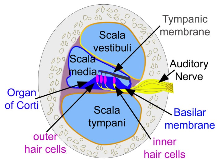
The cochlea is a spiral-shaped structure located in the inner ear. It is responsible for converting sound waves into electrical signals that are sent to the brain. The cochlea is divided into three main parts: the scala vestibuli, the scala tympani, and the scala media.
The scala vestibuli is filled with perilymph, a fluid that is similar to cerebrospinal fluid. The scala tympani is filled with endolymph, a fluid that is high in potassium ions. The scala media is located between the scala vestibuli and the scala tympani.
It is filled with endolymph and contains the sensory cells that are responsible for hearing.
The cochlea is lined with hair cells that are responsible for converting sound waves into electrical signals. These hair cells are arranged in a spiral pattern along the length of the cochlea. The hair cells are connected to the auditory nerve, which sends the electrical signals to the brain.
The brain then interprets these signals and we perceive them as sound.
Cross-sectional Diagram of the Cochlea
A cross-sectional diagram of the cochlea shows the three main parts of the cochlea: the scala vestibuli, the scala tympani, and the scala media. The scala vestibuli is located at the top of the cochlea and is filled with perilymph.
The scala tympani is located at the bottom of the cochlea and is filled with endolymph. The scala media is located between the scala vestibuli and the scala tympani and is filled with endolymph. The sensory cells that are responsible for hearing are located in the scala media.
- The scala vestibuli is lined with Reissner’s membrane.
- The scala tympani is lined with the basilar membrane.
- The scala media is lined with the tectorial membrane.
The basilar membrane is a thin, elastic membrane that separates the scala vestibuli from the scala tympani. The basilar membrane is responsible for the tonotopic organization of the cochlea. The tonotopic organization of the cochlea means that different frequencies of sound are detected at different locations along the basilar membrane.
The tectorial membrane is a gelatinous membrane that lies over the hair cells. The tectorial membrane is attached to the hair cells and moves when sound waves pass through the cochlea. The movement of the tectorial membrane causes the hair cells to bend, which generates electrical signals that are sent to the brain.
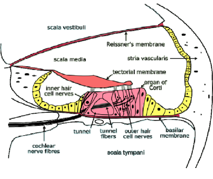
Scala and Membranes

The cochlea is divided into three fluid-filled chambers or scalae: the scala vestibuli, scala media, and scala tympani. These scalae are separated by two membranes: the basilar membrane and the tectorial membrane.
Scala Vestibuli, Cross section of cochlea labeled
The scala vestibuli is the upper chamber of the cochlea. It is filled with perilymph, a fluid that is similar to cerebrospinal fluid. The scala vestibuli is connected to the middle ear by the oval window.
Scala Media
The scala media is the middle chamber of the cochlea. It is filled with endolymph, a fluid that is different from perilymph. The scala media contains the organ of Corti, which is the sensory organ of hearing.
Scala Tympani
The scala tympani is the lower chamber of the cochlea. It is filled with perilymph. The scala tympani is connected to the middle ear by the round window.
Basilar Membrane
The basilar membrane is a thin membrane that separates the scala vestibuli from the scala media. The basilar membrane is wider at the base of the cochlea and narrower at the apex. The width of the basilar membrane determines the frequency of sound that it can detect.
Tectorial Membrane
The tectorial membrane is a thin membrane that overlies the organ of Corti. The tectorial membrane is attached to the tectorial rods, which are cells that are located on the basilar membrane.
Organ of Corti
The organ of Corti is the sensory organ of hearing. It is located in the scala media of the cochlea. The organ of Corti contains hair cells, which are cells that are sensitive to sound. When sound waves reach the cochlea, they cause the basilar membrane to vibrate.
The vibrations of the basilar membrane cause the hair cells to move, which generates electrical signals that are sent to the brain.
Hair Cells and Innervation: Cross Section Of Cochlea Labeled
Hair cells are specialized sensory cells that convert mechanical vibrations into electrical signals, initiating the auditory process. They are located within the organ of Corti, situated on the basilar membrane.
There are two main types of hair cells in the cochlea:
Inner Hair Cells
- More numerous than outer hair cells
- Innervated by afferent nerve fibers from the spiral ganglion
- Transmit auditory information to the brain via the auditory nerve
Outer Hair Cells
- Fewer in number than inner hair cells
- Innervated by efferent nerve fibers from the olivocochlear bundle
- Receive signals from the brain to modulate the stiffness of the basilar membrane, enhancing frequency discrimination
The innervation of the cochlea is provided by the vestibulocochlear nerve (cranial nerve VIII). The afferent fibers from the spiral ganglion transmit auditory information to the cochlear nucleus in the brainstem, while the efferent fibers from the olivocochlear bundle originate in the superior olivary complex and regulate the function of outer hair cells.
Blood Supply and Fluid Dynamics
The cochlea receives its blood supply from the anterior inferior cerebellar artery (AICA). The AICA gives off a labyrinthine artery, which then branches into the cochlear artery. The cochlear artery supplies the cochlea with oxygen and nutrients.Fluid dynamics plays an important role in cochlear function.
The cochlea is filled with fluid, and the movement of this fluid is essential for hearing. When sound waves enter the ear, they cause the eardrum to vibrate. These vibrations are transmitted to the ossicles, which then amplify the vibrations and transmit them to the cochlea.
The vibrations cause the fluid in the cochlea to move, and this movement is detected by the hair cells. The hair cells then send signals to the brain, which interprets the signals as sound.Changes in fluid dynamics can affect hearing.
For example, if the fluid in the cochlea becomes too thick or too thin, it can interfere with the movement of the fluid and cause hearing loss.
Effects of Changes in Fluid Dynamics on Hearing
Changes in fluid dynamics can affect hearing in a number of ways. For example, if the fluid in the cochlea becomes too thick, it can interfere with the movement of the fluid and cause hearing loss. This can occur in conditions such as Meniere’s disease, which is characterized by an increase in the volume and pressure of the fluid in the inner ear.Conversely,
if the fluid in the cochlea becomes too thin, it can also cause hearing loss. This can occur in conditions such as otosclerosis, which is characterized by the formation of a bony growth in the middle ear that can block the transmission of sound waves to the inner ear.In
addition to these conditions, changes in fluid dynamics can also be caused by head trauma or other injuries to the ear. These injuries can damage the delicate structures of the inner ear and lead to hearing loss.
Cross-Sectional Histology
A cross-section of the cochlea reveals a complex and intricate structure that is essential for the sense of hearing. The cochlea is a spiral-shaped organ that is divided into three fluid-filled compartments: the scala vestibuli, the scala media, and the scala tympani.
These compartments are separated by two membranes: the Reissner’s membrane and the basilar membrane.
Scala Vestibuli, Cross section of cochlea labeled
The scala vestibuli is the upper compartment of the cochlea. It is filled with perilymph, a fluid that is similar to cerebrospinal fluid. The scala vestibuli is bounded by the Reissner’s membrane on its medial side and the bony wall of the cochlea on its lateral side.
Scala Media
The scala media is the middle compartment of the cochlea. It is filled with endolymph, a fluid that is high in potassium and low in sodium. The scala media is bounded by the Reissner’s membrane on its lateral side and the basilar membrane on its medial side.
Scala Tympani
The scala tympani is the lower compartment of the cochlea. It is filled with perilymph. The scala tympani is bounded by the basilar membrane on its medial side and the bony wall of the cochlea on its lateral side.
Reissner’s Membrane
The Reissner’s membrane is a thin membrane that separates the scala vestibuli from the scala media. It is composed of collagen and elastin fibers.
Basilar Membrane
The basilar membrane is a thick membrane that separates the scala media from the scala tympani. It is composed of collagen and elastin fibers, and it is supported by the tectorial membrane.
Tectorial Membrane
The tectorial membrane is a gelatinous membrane that lies over the hair cells of the organ of Corti. It is composed of collagen and elastin fibers.
Clinical Significance

Understanding the cross-section of the cochlea is crucial in diagnosing and treating hearing disorders. It provides insights into the intricate anatomy and physiology of the inner ear, enabling clinicians to identify and address pathological changes that affect hearing.
Knowledge of the cochlear cross-section helps in:
- Diagnosing hearing loss:Otoscopy and imaging techniques, such as computed tomography (CT) and magnetic resonance imaging (MRI), allow visualization of the cochlea’s cross-section, revealing structural abnormalities, tumors, or other conditions that may contribute to hearing loss.
- Evaluating cochlear implants:Preoperative imaging of the cochlea’s cross-section helps determine the optimal placement of cochlear implants, ensuring proper stimulation of the auditory nerve and maximizing hearing outcomes.
- Treating Ménière’s disease:Understanding the fluid dynamics within the cochlea’s cross-section is essential for managing Ménière’s disease, a condition characterized by episodes of vertigo, tinnitus, and hearing loss. Treatment options, such as diuretics or surgical decompression, aim to regulate fluid balance and reduce pressure within the cochlea.
Helpful Answers
What is the function of the cochlea?
The cochlea is responsible for converting sound waves into electrical signals that are transmitted to the brain, enabling us to hear.
What are the three main chambers within the cochlea?
The scala vestibuli, scala media, and scala tympani are the three main chambers that run parallel to each other within the cochlea.
What is the role of the basilar membrane in hearing?
The basilar membrane is a flexible structure that vibrates in response to sound waves, stimulating the hair cells that are responsible for converting sound into electrical signals.
